


Written by Dr. Alexandra Koumpi, MD.
(former UK NHS Lead Consultant for Glaucoma)
The following article explains the damage Glaucoma does to our eyes and what causes it. In following articles, I will discuss who is at risk from glaucoma, how we diagnose it, and the current and the future treatments for it.
The Eye Anatomy
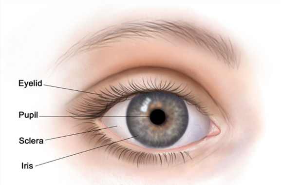
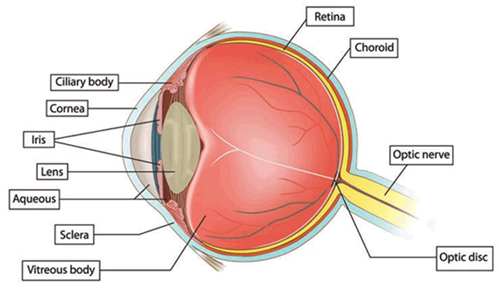
The white Part of the eye is a tough tissue that covers and protects the eye and is called sclera. Only the front part of sclera can be seen. A clear, delicate membrane covers the sclera, called the conjunctiva. At the front of the eye, there is the clear part of the eye’s protective covering, called cornea, which allows light to enter the eye. The colored part of the eye, the iris with a hole in its centre, the pupil, contracts and expands to let just the right amount of light into the eye. The light is directed to the lens which lies behind the pupil.
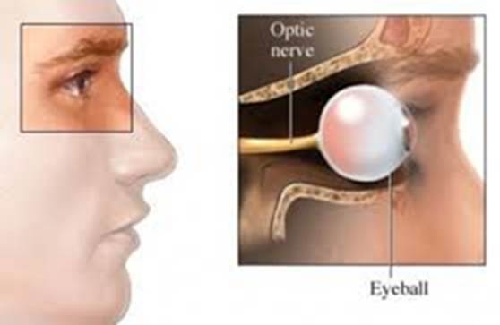
The lens focuses the light onto the retina (lining the back of the eye). Nerve fibers in the retina carry images to the brain through the optic nerve.
What does glaucoma damage?
Glaucoma is a group of eye conditions in which the main nerve to the eye (the optic nerve) is damaged where it leaves the eye (the optic disc).
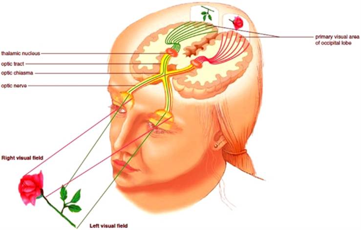
This nerve carries information about what is being seen from the eye to the brain and as it becomes damaged, vision is lost.
This results in misty and patchy vision, with eventual loss of central vision, although this is rare.
Any vision which has been lost to glaucoma cannot be recovered, with early diagnosis, careful monitoring and regular use of treatments, the vast majority of people retain useful sight for life.
The damage to the optic nerve in glaucoma is usually associated with excessive pressure within the eye.
How does this damage happen? – The healthy eye and the healthy drainage
The front part of the eye is filled with a clear fluid called aqueous humor, made by the ciliary body.
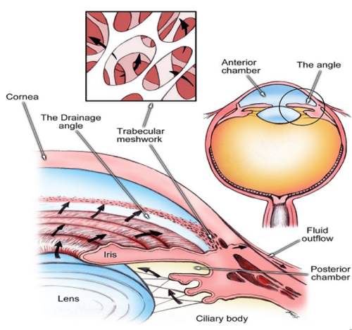
The fluid flows out through the pupil, through the eye’s drainage system. It is then absorbed into the bloodstream
This drainage system is a meshwork of drainage canals around the outer edge of the iris.
Proper drainage helps keep eye pressure at a normal level.
The production, flow, and drainage of this fluid is an active continuous process that is needed for the health of the eye.
The inner pressure of the eye (Intraocular Pressure or IOP) depends upon the amount of fluid in the eye. If your eye’s drainage system is working properly then fluid can drain out and prevent a buildup. Likewise, if your eye’s fluid system is working properly, then the right amount of fluid will be produced for a healthy eye. The IOP can vary at different times of the day, but it normally stays within a range that the eye can handle.
How does this damage happen? – The Eye with Glaucoma
About 1 million small individual thread-like nerve fibers run from the retina to the optic nerve. These fibers meet at the optic disc.
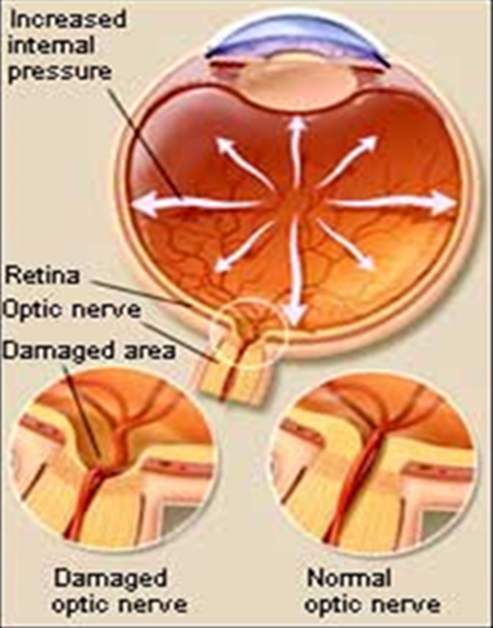
If the drainage of fluid, out of the eye isn’t working as well as it should be, the pressure in the eye (the intraocular pressure) gets too high, it squeezes the optic nerve where it leaves the eye (optic disc), kills some of its fibers and as they die, the disc begins to hollow and develops a cupped or curved shape. If the pressure remains too high for too long, the extra pressure can also reduce the amount of blood that can get through the tiny blood vessels that supply the nerve and eventually damage the optic nerve and result in sight loss.
It was once thought that high intraocular pressure (IOP) was the main cause of this optic nerve damage. Although IOP is clearly a risk factor, we now know that other factors must be involved because people with “normal” IOP can experience vision loss from glaucoma.
Dr. Koumpi practices at 26-28 Arch. Makariou, Polis Chrysochous