


Written by Dr. Alexandra Koumpi, MD. (former UK NHS Lead Consultant for Glaucoma)
In the previous article I explained that when the drainage of fluid, out of the eye isn’t working, the pressure in the eye (the intraocular pressure) gets too high, and it squeezes the optic nerve. This reduces the amount of blood that can get through the tiny blood vessels that supply the optic nerve and eventually damages it, resulting in sight loss. Which poses the question “what causes the drainage of fluid, out of the eye not to work?”
Types of glaucoma
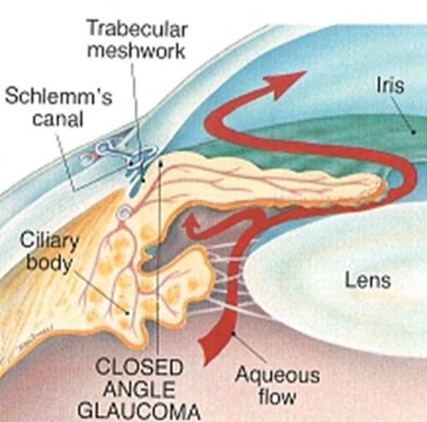
The drainage system is a meshwork of drainage canals (Trabecular meshwork) around the outer edge of the iris. It is located at the drainage angle formed between the iris and cornea. For reasons unknown, an obstruction occurs inside the meshwork of drainage canals despite their entrances being clear and the angle being open. This is known as the Primary Open Angle Glaucoma (POAG), the most common form of glaucoma, accounting for at least 90% of all glaucoma cases. It is also known as chronic open angle glaucoma because it is a chronic (slowly-developing) condition. The eye pressure rises very slowly and there is no pain to warn of a problem, even though the optic nerve is being damaged. When this occurs, part of the field of vision in that eye is damaged. However, the other eye may ‘fill in’ the gap if damage hasn’t occurred in the same part of the field of vision. For this reason, damage has often been done before the person with glaucoma realises there is a problem with his/her sight.

It is also worthwhile to mention the Normal-Tension Glaucoma (NTG) where the optic nerve is damaged even though the eye pressure is not very high. It resembles the POAG.
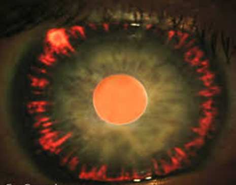
There are further eye conditions that obstruct the drainage canals with different materials within the open angle causing Secondary Open Angle Glaucoma. One such eye condition is the Pseudoexfoliation where a flaky, dandruff-like material peels off the outer layer of the lens within the eye. The material collects in the angle between the cornea and iris and can clog the drainage system of the eye, causing the eye pressure to rise. Patients with this glaucoma show higher pressures and faster disease progression than patients with classic primary open-angle glaucoma.

Another eye condition is Pigment dispersion syndrome where the pigment granules that are in the back of the iris (the colored part of the eye) break into the clear fluid produced inside the eye. These tiny pigment granules flow toward the drainage canals in the eye and slowly clog them, causing the eye pressure to rise. It is estimated that pigment dispersion syndrome develops into pigmentary glaucoma in about 30% of cases and tends to occur at a younger age.
Uveitis is an eye condition that causes swelling and inflammation of the uvea, the middle layer of the eye. Uveitis can occur for a variety of reasons, such as Herpes, Juvenile Idiopathic Arthritis, ankylosing spondylitis and Sarcoidosis. Unfortunately, glaucoma can afflict up to 20% of these patients. The relationship between uveitis and glaucoma is a complex one. Uveitis can cause increased IOP when inflammatory debris obstructs the trabecular meshwork resulting in decreased fluid drainage. In addition, corticosteroid treatment can, by itself, cause elevated IOP as a side effect.
Injury to the eye may cause Traumatic glaucoma. This form of open-angle glaucoma can occur immediately after the injury or develop years later. It can be caused by blunt injuries that bruise the eye (called blunt trauma) or by injuries that penetrate the eye. When a blunt trauma occurs, damage to the drainage system can occur. This can cause bleeding inside the eye. The excess amount of blood, plasma and debris can accumulate and clog the drainage system.

Other eye conditions that lead to the development of secondary open angle glaucoma are the Hypermature cataract, the Thyroid Eye disease, the Sturge Weber syndrome and more.
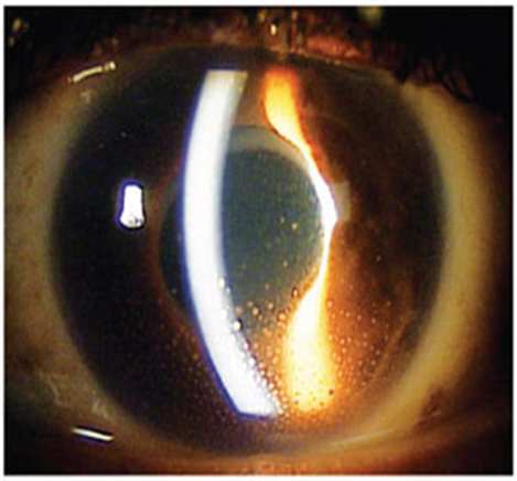
When the drainage angle is structurally narrow or suddenly closes then Primary Angle Close Glaucoma developsvery quickly. The narrow angle is common in longsighted patients. Under certain conditions the sudden closure of the angle with eye pain can occur. This is an emergency that demands immediate medical attention.
When eye conditions such as Uveitis, New vessels (as in Diabetes or Trauma), cataract cause secondarily closure of the drainage angle then the Secondary Angle Close Glaucoma develops.
It is important to diagnose the type of glaucoma in order to provide the right care.
The examination tools we use for the right diagnosis will be presented in a following article.
Dr. Koumpi practices at 26-28 Arch. Makariou, Polis Chrysochous www.akeyecare.com